CHIDAMBARAM HOSPITAL
चिदंबरम अस्पताल,
ചിദംബരം ഹോസ്പിറ്റൽ
சிதம்பரம் மருத்துவமனை,
திசையன்விளை.627657
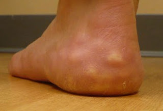
Piezogenic pedal papules are lesions that appear on the feet and wrists.
These can either be accompanied by pain or be asymptomatic and they
usually take the form of papules. It seems that they appear when fat
herniates through the dermis, having a characteristic popular
appearance. This is a common medical condition and generally
asymptomatic in the majority of people. It is not genetically inherited
and it is not related in general to connective tissue disease (in
certain cases, it might be associated with such conditions but this is
not a direct cause). As for the actual papules, these become obvious
when the person stands up, distributing the entire body weight on the
heels.
Symptoms of Piezogenic Pedal Papules
These are the most common symptoms of the piezogenic pedal papules:
- Skin lesions in the form of papules appear on the feet and wrists
- Pale to skin-colored
- Obvious when weight is distributed on the feet (standing up), disappear when the person sits down and the weight is reduced
- Bilateral involvement
- Most often affects the heel of the foot, including the medial, posterior and lateral side
- In the wrist, the volar surface is the one most affected (because of the pressure)
- Pain might accompany the papules, preventing the person from performing certain activities
- The papules present on the feet or wrists can be easily compressed
What are the Causes of Piezogenic Pedal Papules?
These are the most common causes associated with the appearance of the piezogenic pedal papules:
- Excessive weight – obese patients have more weight to bear on their
feet and this is why they present an increased risk for fat to herniate
through the dermis layer of the skin
- Occupation
- Standing for prolonged periods of time
- Constant pressure applied to the wrist
- Associated with Ehlers-Danlos syndrome (collagen disorder)
- Idiopathic (unknown cause)
- Orthopedic problems
- Can occur in newborns (no predisposition)
- Excessive weight bearing (physical exercise or other vigorous physical activity)
- Repetitive pressure force on the said areas
Diagnosis
These are the most common methods used for the diagnosis of piezogenic pedal papules:
- Clinical examination
- Identification of fat herniating through the dermis
- No laboratory analysis
- No imaging studies
- Differential diagnosis – this can be made with the following medical conditions:
- Infantile pedal papules (bilateral congenital adipose plantar
nodules/precalcaneal congenital fibrolipomatous hamartomas/pedal
papules) – appear in newborns
- Xanthomas – painful lesion that occurs most often on the buttocks but can affect other areas of the body as well
- Tophi – appear in patients with gout, being represented by solid urate that deposits in the connective tissue.
Treatment
These are the most common treatment courses and recommendations made for people diagnosed with piezogenic pedal papules:
- No oral or topical medication available or necessary
- Orthotics and other devices are recommended in patients who exhibit symptoms
- Supportive external pressure device
- Heel taping
- Compression stockings
- Foam rubber foot pads
- Foam-fitting plastic heel cups
- Rest and elevation might help with the symptoms (temporary relief)
- Non-surgical approach
- Injections with betamethasone and bupivacaine – these are
recommended for patients who have been diagnosed with Ehlers-Danlos
syndrome, reducing the painful symptoms. Repeated injections are
necessary for complete pain relief.
- No surgery has proven to be effective for the treatment of
piezogenic pedal papules but the surgical approach can be recommended
for a skin lesion that is persistent and intensely painful.
- Avoiding periods of prolonged standing, walking or running can help
if the papules are painful (practically, the patient is recommended
avoid any kind of physical activity that might involve excessive or
prolonged weight bearing)
- In case of excessive weight, patients are recommended to follow a weight loss program
- Recent studies recommend electro-acupuncture as treatment for this medical condition.


CHIDAMBARAM HOSPITAL
चिदंबरम अस्पताल,
ചിദംബരം ഹോസ്പിറ്റൽ
சிதம்பரம் மருத்துவமனை,
திசையன்விளை.627657
- தீவிர சிகிச்சை மருத்தவம்
- பொது மருத்துவரம்
- பொது அறுவை சிகிச்சை
- குழந்தை அறுவை சிகிச்சை
- குழந்தை லேப்ராஸ்கோப்பி அறுவை சிகிச்சை
- Cesarean section
- Dilation and Curettage
- Vulvectomy
- Tubal Ligation
- Trachelectomy
- Selective Salpingography
- Myomectomy
- Hysterosalpingography
-Endometrial or Uterine Biopsy
- Colporrhaphy
-Vaginal hystectomy
- Appendicitis
- Lymphangioma
- Cleft lip and palate
- Esophageal atresia and tracheoesophageal fistula
- Hypertrophic pyloric stenosis
- Intestinal atresia
- Necrotizing enterocolitis
- Imperforate anus
- Undescended testes
- Omphalocele
- Gastroschisis
- Hernias
- Teratomas
- Amputation
- Appendectomy
- Cholecystectomy
- Colectomy
- Cystoscopy
- Hemorrhoidectomy
- Hysterectomy
- Hysteroscopy
- Inguinal Hernia
- Laparoscopy
- Mastectomy
- Thyroidectomy
- Tracheostomy
- Tonsillectomy and Adenoidectomy
- Umbilical Hernia
- லேப்ராஸ்கோப்பி அறுவை சிகிச்சை
- மகப்பேறு மருத்துவம்
- தாய்மை மருத்துவம்
- மகளிர் நோய் இயல்
- சர்க்கரை வியாதி மருத்தவம்
- X - ரே (X-Ray)
- ஈசிஜி (ECG)
- இரத்த ஆய்வு (Blood Investigation LAB)
- அல்ட்ராசவுண்ட் ஸ்கேன்
(ULTRASOUNDSCAN)
- பிசியோதெரபி பயிற்சி (PHYSIOTHERAPY)
- முக வாதம் தூண்டுதல் பயிற்சி (BELLS PALSY STIMULATION)
- துரக்கம்-முதுகு வலி நிவாரணத் பயிற்சி(TRACTION)
- மெழுகு ஓத்தLம் (WAX BATH)
- அகச்சிவப்பு கதிர் வலி நிவாரணத் ஓத்தLம்(INFRA RED Hot Fermentation)
Dr.M.I. கிறிஸ்டோபர் சாமுவேல் MBBS,MS.,FIAGES.,லேப்ராஸ்கோப்பி அறுவை சிகிச்சை நிபுணர்.,
DR.அலெக்ஸ் J கிறிஸ்டோபர் MBBS,MS,MCH.,(PAEDIATRIC SURGEON),லேப்ராஸ்கோப்பி அறுவை சிகிச்சை நிபுணர்.,
DR.அருண் G கிறிஸ்டோபர் MBBS,MD(Anaesthesia)மயக்க மருந்து நிபுணர்,Pain Management., Dip.Diab., சர்க்கரை வியாதி மருத்துவர்.,
PT.அந்தோணி றீகன் B.P.T
(பிசியோதெரபி நிபுணர்)MCSE,COPA,D.Pharm.,


Comments
Post a Comment